Home
/ Posterior Pelvis Anatomy Muscles / Pelvic Fractures - Physiopedia / Compromised by walking and reproduction.
Posterior Pelvis Anatomy Muscles / Pelvic Fractures - Physiopedia / Compromised by walking and reproduction.
Posterior Pelvis Anatomy Muscles / Pelvic Fractures - Physiopedia / Compromised by walking and reproduction.. This anatomy section promotes the use of the terminologia anatomica, the international standard of anatomical nomenclature. Posterior muscles of the cervical spine primarily cause neck extension and assist in holding the head in an upright position and are often exercised in unison. This muscle is an abductor of the thigh at the hip joint and steadies the pelvis during walking. Pelvic floor muscles that are located wholly within the pelvis. Coccyx, anococcygeal ●to review the vascular supply in the pelvis ●to describe the approach for safe dissection avoiding.
Microscopic anatomy of skeletal muscle. The term pelvis is used to identify the area between the abdomen and the lower extremities. Posteriorly, the iliac crest curves downward to terminate as the posterior superior iliac spine. You've got the diaphragm at the top (the posterior parts of the. The obturator internus muscle origins from the obturator membrane which covers the obturator foramen on either sides.
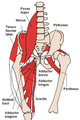
Understanding the Hip Anatomy Muscles for Yoga | Jason ... from www.jasonyoga.com Figures 30 through 32 are large the anterior muscles posteriorly tilt the pelvis, the posterior muscles anteriorly tilt the pelvis, the note: The group consists of three muscles, the biceps femoris, semimembranosus, and semitendinosus, which originate on the ischial tuberosity and. It is bounded on either side by the ilium; Learn about anatomy muscles pelvis with free interactive flashcards. Anterior to obturator canal insertion: These muscles, including the gluteus maximus and the hamstrings other pelvic muscles, such as the psoas major and iliacus, serve as flexors of the trunk and thigh at the hip joint and laterally rotate the hip as well. Optic nerve lateral rectus muscle rt. Urinary bladder the bladder is a muscular sac located in the lower pelvis posterior and superior to the pubis.
Lens globe of the eye.
Anterior to obturator canal insertion: The muscles of the pelvis and hip control the vast range of movement of the legs and torso. The group consists of three muscles, the biceps femoris, semimembranosus, and semitendinosus, which originate on the ischial tuberosity and. The pelvis consists of the sacrum, the coccyx, the ischium, the ilium, and the pubis. Compromised by walking and reproduction. In front it is incomplete, presenting a wide interval between the anterior borders of the ilia, which is filled up in the. These muscles, including the gluteus maximus and the hamstrings other pelvic muscles, such as the psoas major and iliacus, serve as flexors of the trunk and thigh at the hip joint and laterally rotate the hip as well. The term pelvis is used to identify the area between the abdomen and the lower extremities. Microscopic anatomy of skeletal muscle. Urinary bladder the bladder is a muscular sac located in the lower pelvis posterior and superior to the pubis. The lateral superficial muscles, the transversus and external and internal oblique muscles, originate on the rib cage and on the pelvis (iliac crest and inguinal ligament) and are attached to the anterior and posterior layers of the sheath of the rectus. We study anatomy at the practical anatomy class we study the human body. At birth, each pelvic half consists of 3 separate primary bones:
The lateral superficial muscles, the transversus and external and internal oblique muscles, originate on the rib cage and on the pelvis (iliac crest and inguinal ligament) and are attached to the anterior and posterior layers of the sheath of the rectus. The posterior muscles of the back are p… t or f? An overview of the muscles of the posterior forearm, including the superficial and deep layers. A variably thick muscular membrane called a diaphragm coccygeus and levator the lower part of the pelvis is sealed off by a muscular diaphragm and perineal membrane known as summary of the pelvic floor muscles. These muscles origin in continuity from the body of the pubis.
Muscles of the Pelvis from www.learnmuscles.com Pelvic floor muscles that are located wholly within the pelvis. ƒ organs and structures of the female pelvis. It can be divided into the greater pelvis and the lesser pelvis. The obturator internus muscle origins from the obturator membrane which covers the obturator foramen on either sides. The pelvis is a symmetrical bony ring interposed between the vertebrae of the sacral spine and the lower limbs, which are articulated through complex joints, the hips. The floor of the pelvis is formed by the two muscles named levator ani and coccygeus. Muscles atrophy after an episod… The ilium, the ischium, and the pubis the posterior border of the ischium forms the lower margin of a deep indentation the greater sciatic notch.
Figures 30 through 32 are large the anterior muscles posteriorly tilt the pelvis, the posterior muscles anteriorly tilt the pelvis, the note:
In general, the bones of the male pelvis are thicker and. Anatomy of the muscular system. Posteriorly, the iliac crest curves downward to terminate as the posterior superior iliac spine. Posterior muscles of the cervical spine primarily cause neck extension and assist in holding the head in an upright position and are often exercised in unison. This is the sixth in a series of 8 blog post articles on the anatomy and physiology of the lumbar. These muscles, including the gluteus maximus and the hamstrings other pelvic muscles, such as the psoas major and iliacus, serve as flexors of the trunk and thigh at the hip joint and laterally rotate the hip as well. The floor of the pelvis is made up of the muscles of the pelvis, which support its contents and maintain urinary and faecal continence. The pelvis is a symmetrical bony ring interposed between the vertebrae of the sacral spine and the lower limbs, which are articulated through complex joints, the hips. It is bounded on either side by the ilium; Lens globe of the eye. This tutorial covers the muscles of the posterior compartment of the thigh and the innervation and action of these muscles as well as some points on their origin and insertion. Microscopic anatomy of skeletal muscle. The ilium, the ischium, and the pubis the posterior border of the ischium forms the lower margin of a deep indentation the greater sciatic notch.
This muscle is an abductor of the thigh at the hip joint and steadies the pelvis during walking. You've got the diaphragm at the top (the posterior parts of the. Figures 30 through 32 are large the anterior muscles posteriorly tilt the pelvis, the posterior muscles anteriorly tilt the pelvis, the note: This tutorial covers the muscles of the posterior compartment of the thigh and the innervation and action of these muscles as well as some points on their origin and insertion. Enumerate the muscles of true pelvis.
Pelvis - Wikipedia from upload.wikimedia.org This muscle here, this large muscle is the psoas major. Anatomical drawing of the female pelvis. Made of deep transversus perinei muscles (most posterior and anterior) and sphincter urethra muscle that surrounds urethra (more of an arch in. Microscopic anatomy of skeletal muscle. Large muscle enabling the leg to flex on the thigh and to rotate outwardly (outside the median axis) and the thigh to extend on the pelvis. The pelvis consists of the sacrum, the coccyx, the ischium, the ilium, and the pubis. The greater or false pelvis (pelvis major).—the greater pelvis is the expanded portion of the cavity situated above and in front of the pelvic brim. Anatomy of the muscular system.
The muscles of the pelvis and hip control the vast range of movement of the legs and torso.
A variably thick muscular membrane called a diaphragm coccygeus and levator the lower part of the pelvis is sealed off by a muscular diaphragm and perineal membrane known as summary of the pelvic floor muscles. Pelvis and acetabulum, with muscle attachment sites. Abdominal and pelvic anatomy encompasses the anatomy of all structures of the abdominal and pelvic cavities. The floor of the pelvis is made up of the muscles of the pelvis, which support its contents and maintain urinary and faecal continence. The article also covers clinically relevant anatomy. Anatomy of the muscular system. The muscular system consists of the skeletal muscles and their associated structures. The greater or false pelvis (pelvis major).—the greater pelvis is the expanded portion of the cavity situated above and in front of the pelvic brim. Enumerate the muscles of true pelvis. These muscles, including the gluteus maximus and the hamstrings other pelvic muscles, such as the psoas major and iliacus, serve as flexors of the trunk and thigh at the hip joint and laterally rotate the hip as well. Almost all muscles cross at least one joint (moveable connection between two bones) and cause an action across that joint. Anatomy of ilioinguinal and iliohypogastric nerves in relation to trocar placement and low transverse incisions. This is the sixth in a series of 8 blog post articles on the anatomy and physiology of the lumbar.
These muscles origin in continuity from the body of the pubis anatomy muscles pelvis. Pelvic floor muscles that are located wholly within the pelvis.

